Published on jan 4 2020 this video provides a clear and practical introduction to chest xray. Chest x ray interpretation tubes and lines duration. Chest drains are usually inserted through the chest wall in the mid axillary line.
chest tube x ray pneumothorax chest tube x ray pigtail chest tube x ray baby chest x ray tube tracheostomy tube chest x ray

This x ray was performed to check the position of a chest drain used to treat a pneumothorax conventionally this x ray would be considered inadequate for diagnostic purposes as it does not include the costophrenic angles the chest drain position is shown clearly the image does not need to be repeated to include the costophrenic angles.
Chest tube x ray. Lines and tubes are important components in chest radiographic evaluation. Chest x rays are commonly used to confirm correct positioning of certain medical devices and to check for associated complications following placement or misplacement. The focus is on developing a simple but still detailed approach. Assessment of their position is important but they also give you an idea about how sick the patient is and narrow down the differential diagnosis.
We have a more in depth reference article see medical devices in the. Below the left hemidiaphragm. Chest drain treatment for pneumothorax to drain a pneumothorax the tube is aimed superiorly towards the apex of the pleural cavity. The video is designed for physicians.
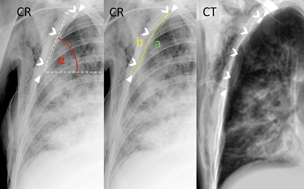
Chest x ray lines and tubes can be easily assessed and should be the first thing that you look at when reviewing a chest x ray. The superiorinferior and mediallateral positioning of the tube can be determined on a chest x ray. Chest x ray interpretation et tube duration. This tutorial describes the correct anatomical location following placement of common tube devices seen on chest x rays.
Pharyngeal or esophageal perforation. This is a summary article. Insertion into trachea or bronchus pneumoniapulmonary contusionpulmonary laceration. Ng tube tip 10 cm distal to the gastro esophageal junction.